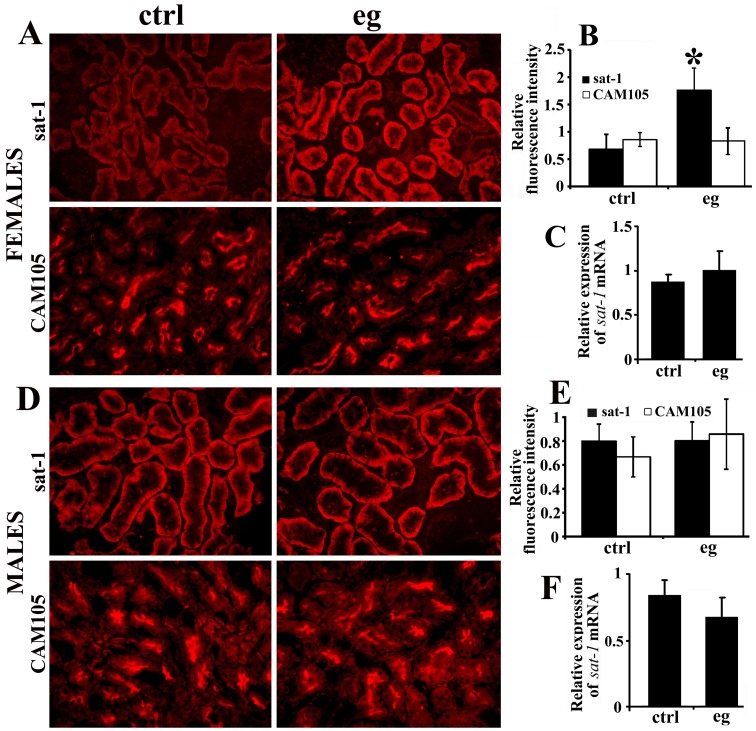
Figure 4.
Sulfate anion transporter sat-1 (Slc26a1) and cell adhesion molecule 105 (CAM105) protein expression (A, B, D, E) and sat-1 mRNA expression (C, F) in the kidneys of control (ctrl) and EG-treated (eg) rats. (A-C) Data in female rats. (A) Immunostaining in tissue cryosections. In EG-treated females the intensity of sat-1-related staining of the basolateral membrane in cortical proximal tubules was stronger than that in controls, whereas the intensity of CAM105-related staining, localized to the proximal tubule brush-border membrane, was similar. (B) Relative fluorescence pixel intensity showed ~ 3-fold increase in the sat-1-related staining intensity in EG-treated females (*vs control, P < 0.001; n = 6 in each group) and no change for CAM105 (P = 0.731; n = 6 in each group). (C) Relative expression of sat-1 mRNA was similar in control and EG-treated female rats (P = 0.223; n = 6 in each group). (D-F) Data in male rats. (D) Immunostaining in tissue cryosections. In EG-treated males the intensity of sat-1-related staining of the basolateral membrane in cortical proximal tubules, and the CAM105-related staining in the proximal tubule brush-border membrane, was similar to that in controls. (E) Relative fluorescence pixel intensity showed no differences in the sat-1-related (P = 0.927) and CAM105-related (P = 0.220) staining intensity between the control and EG-treated males (n = 6 in each group). (F) Relative expression of sat-1 mRNA was similar in control and EG-treated male rats (P = 0.125; n = 6 in each group). Bar, 20 μm.

