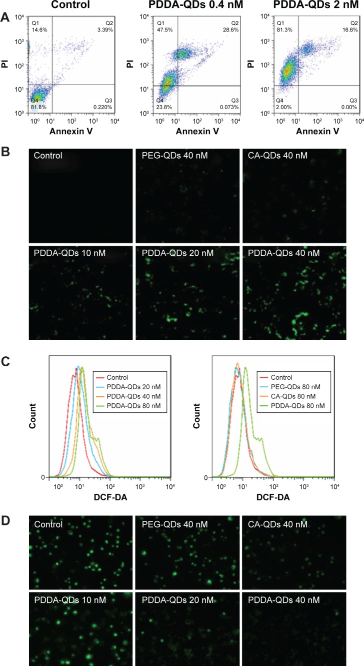
Figure 6.
The underlying mechanisms of QDs cytotoxicity.
Notes: Apoptotic or necrotic cell death (A) of MDA-MB-231 cells was detected by flow cytometry after 24-hour incubation with PDDA-QDs, followed by Annexin V-FITC and PI dual-staining. Intracellular reactive oxygen species (ROS) production in MDA-MB-231 cells treated with different charged QDs was detected by confocal microscopy (B) and flow cytometry (C), respectively. Intracellular ROS was measured using 10 µM DCF-DA probe. (D) The membrane mitochondria potential of MDA-MB-231 cells was acquired with fluorescence microscopy. Cells were incubated with various concentrations of QDs for 2 hours, and treated with 40 nM of DiOC6(3) for 30 minutes.
Abbreviations: CA, carboxylic acid; DCF-DA, 2′,7′-Dichlorofluorescin diacetate; FITC, fluorescein isothiocyanate; PDDA, polydiallydimethylammounium chloride; PI, propidium iodide; PEG, polyethylene glycol; QDs, quantum dots.

