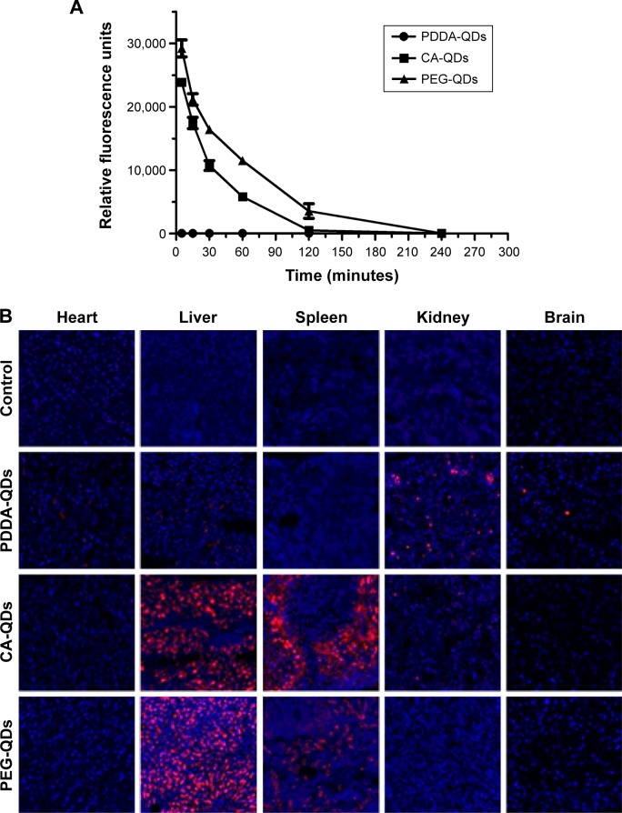
Figure 7.
In vivo blood clearance and tissue biodistribution.
Notes: In vivo blood clearance (A) and tissue biodistribution (B) of different charged QDs in BALB/c mice after intravenous administration. QDs in the serum were detected by the fluorescence measurement using NanoDrop Fluorospectrometer. The major organs were excised for microscopic examination at 4 hours postinjection. The organ slices (10 µm) were prepared with a cryostat, air-dried for 30 minutes, and fixed with 4% paraformaldehyde for 10 minutes. The nuclei were stained by DAPI (blue), and the signal of QDs (red) was acquired with confocal fluorescence microscopy. Magnification ×200.
Abbreviations: CA, carboxylic acid; DAPI, 4′,6-diamidino-2-phenylindole; PDDA, polydiallydimethylammounium chloride; PEG, polyethylene glycol; QDs, quantum dots.

