
Figure 1.
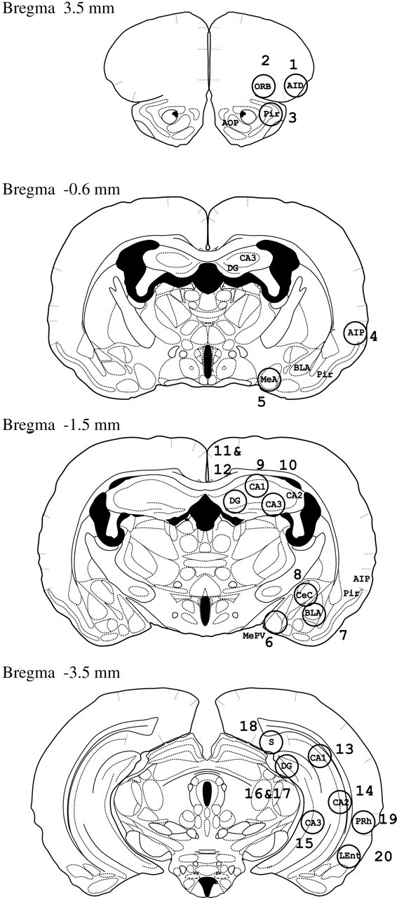
Locations (shown by numbered circles) of the 20 brain areas that we examined in four sections at different locations relative to Bregma, based on the hamster brain atlas of Morin and Wood (2001). The 20 brain areas and abbreviations are as follows: (1) AID, agranular insular cortex, anterior; (2) ORB, orbital cortex; (3) Pir, piriform cortex; (4) AIP, agranular insular cortex, posterior; (5) MeA, medial amygdala; (6) MePV, medial amygdala, posteroventral; (7) BLA, basolateral amygdala; (8) CeC, central amygdala nucleus, capsular; (9) ADHCA1, CA1 region of anterior dorsal hippocampus; (10) ADHCA3; (11) ADHCA4; (12) ADHDG, dentate gyrus of anterior dorsal hippocampus; (13) PDHCA1, CA1 region of posterior dorsal hippocampus; (14) PDHCA2;(15)PVHCA3, CA3 region in posterior ventral hippocampus; (16)PDHCA4; (17)PDHDG, dentate gyrus of posterior dorsal hippocampus; (18) PDS, posterior dorsal subiculum; (19) PRh, perirhinal cortex; and (20) LEnt, lateral entorhinal cortex.