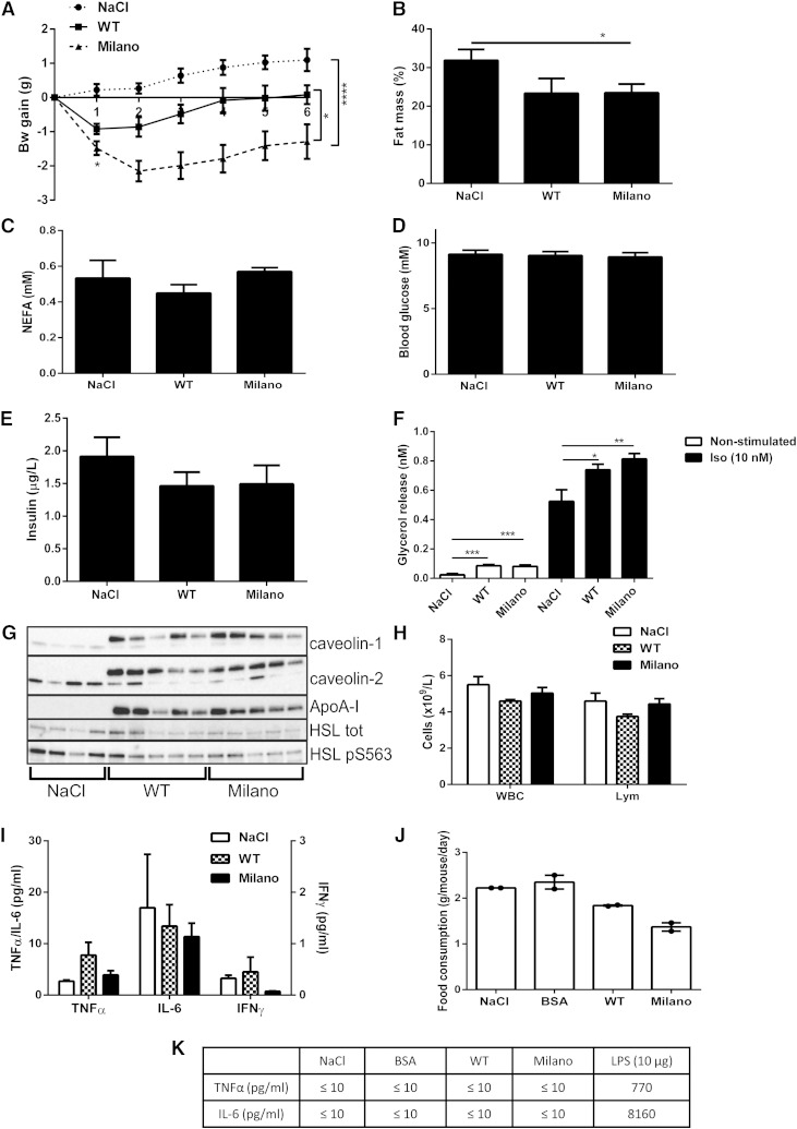
Fig. 1.
ApoA-I Milano treatment induces rapid weight loss and increased lipolysis. C57bl6/J mice received one intraperitoneal injection/day of ApoA-I Milano (20 mg/kg), WT (20 mg/kg), or saline (NaCl), during 6 days (n = 6–11 animals/group). Body weight was monitored daily during the treatment and 24 h after the last injection (day 0 first injection, day 5 last injection) (A). Data presented in B–I were collected 24 h after the last treatment (n = 6–11 animals/group); fat mass determined by DEXA body scan (% body fat mass) (B), serum NEFA (mM) (C), blood glucose (mM) (D), and serum insulin (µg/l) (E). Adipose cells were isolated from epididymal fat tissue, and lipolysis was measured by analyzing glycerol release into medium, either nonstimulated or isoproterenol stimulated (10 nM, 30 min). (F). Adipose tissue was collected and subjected to Western blot analysis to determine caveolin-1, caveolin-2, and HSL protein expression levels; phosphorylation of HSL pS563; and presence of ApoA-I Milano using an ApoA-I-specific antibody (n = 4–5 animals/group) (G). Whole blood was analyzed to determine WBC count and lymphocytes (Lym) (×109/l) (H). Serum collected the day after last treatment was analyzed for levels of TNF-α, IL-6 (left y-axis, pg/ml), and IFN-γ (right y-axis, pg/ml) (I). Food consumption (g/mouse/day) was measured per cage (5 animals/cage). Data displayed as mean from 2 days (J). Serum collected 24 h after the first injection was analyzed for levels of TNF-α and IL-6 (pg/mol); serum from mice treated with 10 µg lipopolysaccharide for 90 min was used as a positive control (n = 5 animals/group) (K). Data in A–K are presented as mean ± SEM. * P ≤ 0.05, ** P ≤ 0.01, *** P ≤ 0.0005, and **** P ≤ 0.0001.

