
Fig. 1.
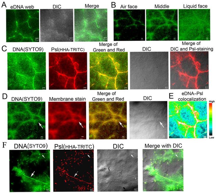
The web of DNA fibres and its association with Psl polysaccharide and bacterial cell membrane in the air–liquid interface biofilms (pellicles) of P. aeruginosa.
A. A web of fibre-like DNA (stained in green by SYTO9) was clearly visualized at the middle of pellicles grown at air–liquid interface of standing culture.
B. The optical sectioned images showed the location of eDNA fibres web (green) in a pellicle.
C. The eDNA fibres web (green) was associated with the fibres of Psl polysaccharide (stained in red by lectin HHA-TRITC) in pellicles.
D. The eDNA fibres were colocalized with bacterial membrane in a 46 h pellicle stained by SYTO9 and the cell membrane stained by FM6-64 (the arrow indicated the eDNA-membrane fibres web).
E. The eDNA–Psl colocalization was mostly associated with the fibre-like matrix structure depicted by the colour map, which was made by the ImageJ software according to the colour image series of (C).
F. The eDNA–Psl fibres with strong (indicated by a big arrow) or weak (indicated by a small arrow) fluorescent signal both found in the middle of a pellicle. Scale bar in all panels: 5 μm.