
Fig. 3.
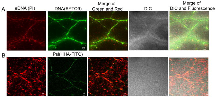
The eDNA and dead bacteria in pellicles of P. aeruginosa PAO1 were stained by Propidium iodide (PI) to confirm that fibre-like DNA was eDNA. Shown were the optical sectioned images in the middle of pellicles. (A) PI (red) and SYTO9 (green) double-stained images of a 2-day-old pellicle. (B) PI (red) and HHA-FITC (a lectin that stains Psl in green) double-stained images of a 2-day-old pellicle. Scale bar: 10 μm for (A); 5 μm for (B).