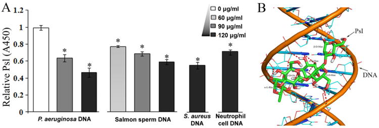
Fig. 5.

The interaction between DNA and Psl polysaccharide.
A. The detection of Psl can be interfered by DNA isolated from P. aeruginosa, salmon sperm, S. aureus and neutrophil cells. Psl was detected by anti-Psl antibody in ELISA assay. The samples without DNA blocking were normalized to 1 (A450 = 0.635 ± 0.012). *P< 0.01.
B. A plausible model for DNA–Psl interaction. The docking result suggested that hydrogen bonds (indicated by dash lines) can be formed between a Psl polysaccharide repeat unit (carbon, green; oxygen, red; hydrogen, white) and the standard B-DNA duplex (phosphorus, orange; carbon, cyan; oxygen, red; nitrogen, blue).
