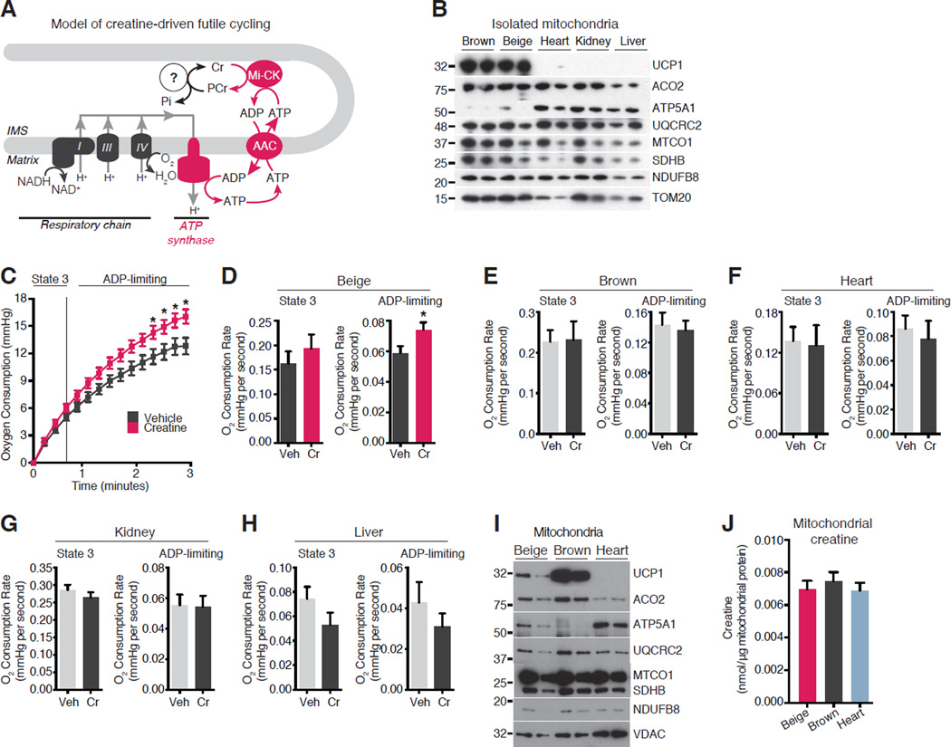
Figure 2. Creatine Stimulates Respiration in Beige Fat Mitochondria When ADP is Limiting.
(A) Model of creatine-based substrate cycling. IMS, intermembrane space; AAC, ADP/ATP Carrier.
(B) Western blot of mitochondrial proteins. N = 2 mitochondrial preparations, 15 mice per cohort.
(C) Oxygen consumption by beige fat mitochondria treated with and without creatine (0.01 mM) in the presence of 0.2 mM ADP. Vertical dashed line, State 3 to State 4 transition. N = 9 mitochondrial preparations, 15 mice per cohort.
(D) State 3 and ADP-limiting Oxygen consumption rate (OCR) of beige fat mitochondria treated with and without creatine in the presence of 0.2 mM ADP. N = 9 mitochondrial preparations, 15 mice per cohort.
(E) State 3 and ADP-limiting OCR of brown fat mitochondria treated as in D. N = 3 mitochondrial preparations, 15 mice per cohort.
(F) State 3 and ADP-limiting OCR of heart mitochondria treated as in D. N = 3 mitochondrial preparations, 8 mice per cohort.
(G) State 3 and ADP-limiting OCR of kidney mitochondria treated as in D. N = 3 mitochondrial preparations, 15 mice per cohort.
(H) State 3 and ADP-limiting OCR of liver mitochondria treated as in D. N = 3 mitochondrial preparations, 2 mice per cohort.
(I) Western blot of mitochondrial proteins from beige fat, brown fat, and heart. N = 2 mitochondrial preparations, 15 mice per cohort.
(J) Mitochondrial creatine concentration in beige fat, brown fat, and heart. Data are presented as mean ± SEM. *p < 0.05.

