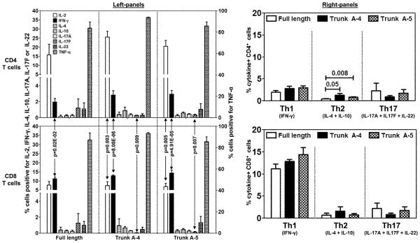
Figure 7. Cytokine responses to dual epitopes of FL-Myhc334-352 in CD4 and CD8 T cells.
LNCs obtained from A/J mice immunized with Myhc334-352, Trunk A-4, Trunk A-5 or Trunk A-6 were stimulated with the immunizing peptides for two days, and the cultures were maintained in IL-2 medium. Cells were briefly stimulated on day 6 with PMA and ionomycin for 5 hours in the presence of monensin. After staining with anti-CD4, anti-CD8 and 7-AAD, cells were fixed and permeabilized, and then stained with cytokine antibodies or their respective isotype controls. Cells were acquired by flow cytometry and the percentages of cytokine-producing cells in the live (7-AAD−) CD4 or CD8 T cell subsets were determined using Flow Jo software in relation to the gates drawn for isotype controls corresponding to each cytokine. Mean ± SEM values from four to six independent experiments are shown. In the left panel, cells positive for individual cytokines are shown (left, y-axis: IL-2, IFN-γ, IL-4, IL-10, IL-17A, IL-17F and IL-22; right, y-axis: TNF-α). The right panel shows the cytokine+ cells with respect to Th1 (IFN-γ), Th2 (IL-4 + IL-10) and Th17 (IL-17A + IL-17F + IL-22) subsets.

