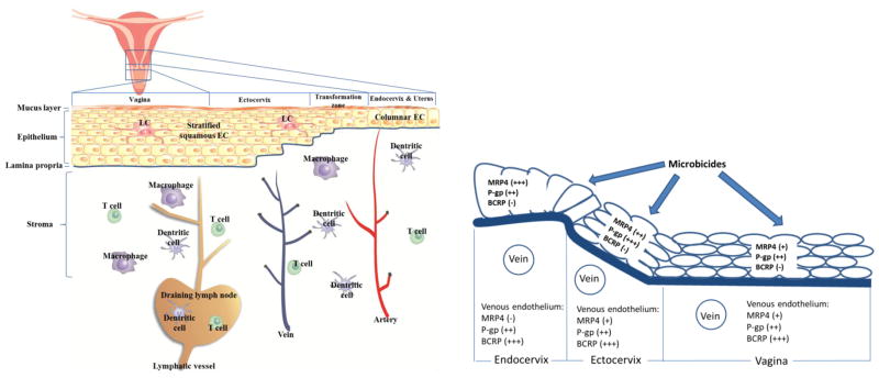
Figure 1. Anatomy and transporter expression in the female genital tract.
The FGT that are potential sites of sexual HIV transmission include uterus, endocervix, ectocervix, and vagina. A mucus layer covers the epithelia of all these tissue segments, serving as a physical barrier to vaginally administered drugs. Stratified, squamous epithelial layers line the vagina and ectocervix, and a single-layer of columnar epithelial cells lines the endocervix and uterus. The epithelial layers and stroma are separated by the collagen-rich laminar propria. The CD4+ T cells, dentritic cells and macrophages are HIV target cells, and they are distributed in epithelial layers, stroma, and draining lymph nodes. The invading HIV particles can infect these immune cells and establish local tissue infection, expansion, and progress to systemic dissemination. Blood vessels (veins, arteries) and lymphatic vessels mediate the distribution of drug between the tissues and systemic compartments (circulating blood and lymph). LCs, Langerhan’s cells, which are the dentritic cells residing in peripheral tissues. EC, epithelial cells. (The figure is based on Zhou et al. [62])

