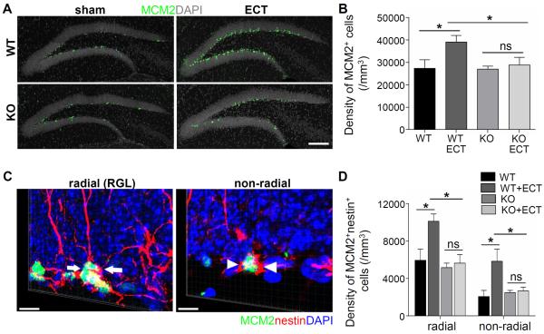
Figure 1. Crucial role of Gadd45b in electroconvulsive shock (ECS)-induced proliferation of quiescent radial glia-like (RGLs) neural stem cells and non-RGL neural precursors.
(A–B) Gadd45b mediates ECS-induced proliferation of neural stem cells in the subgranular zone (SGZ). A. Representative confocal images of immunostaining of MCM2 (green), an endogenous cell proliferation marker, and DAPI staining (grey) in the dentate gyrus of adult Gadd45b knockout (KO) and wild-type (WT) littermates with or without ECS. Scale bar: 50 μm. B. Summary of stereological quantification of MCM2+ cells in the adult dentate gyrus of WT and KO littermates with or without ECS. Values represent mean ± SEM (n = 4 animals per group; *P < 0.05, one-way ANOVA; ns, no significance). (C–D) Gadd45b mediates ECS-induced activation of RGL and non-RGL neural stem cells in the SGZ. C. Sample confocal images of immunostaining of MCM2 (green), nestin (red) and DAPI staining (blue). Scale bars: 25 μm. Arrows point to MCM2+nestin+ RGL (left; arrows) and MCM2+nestin+ non-RGL neural precursors (right; arrowheads), respectively (See Supplementary Movie 1). D. Summary of stereological quantification of MCM2+nestin+ RGL and non-RGL precursors in the adult dentate gyrus of WT and KO littermates with or without ECS. Values represent mean ± SEM (n = 4 animals per group; *: P < 0.05, one-way ANOVA; ns, no significance).

