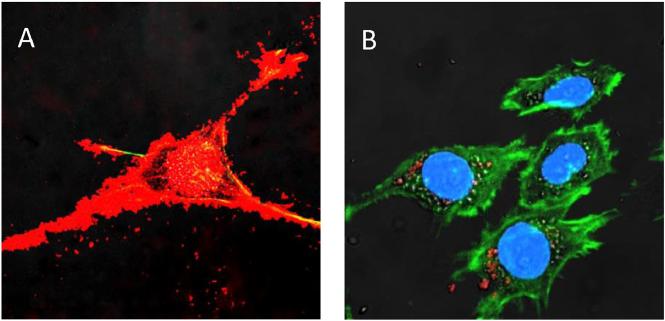
Fig. 1.
A profound accumulation of exosomes (A, red) compared to polymer-based nanoparticles (B, red) in target PC12 neuronal cells stained for actin microfilaments (green) and nuclei (blue). (For interpretation of the references to color in this figure legend, the reader is referred to the web version of this article.)

