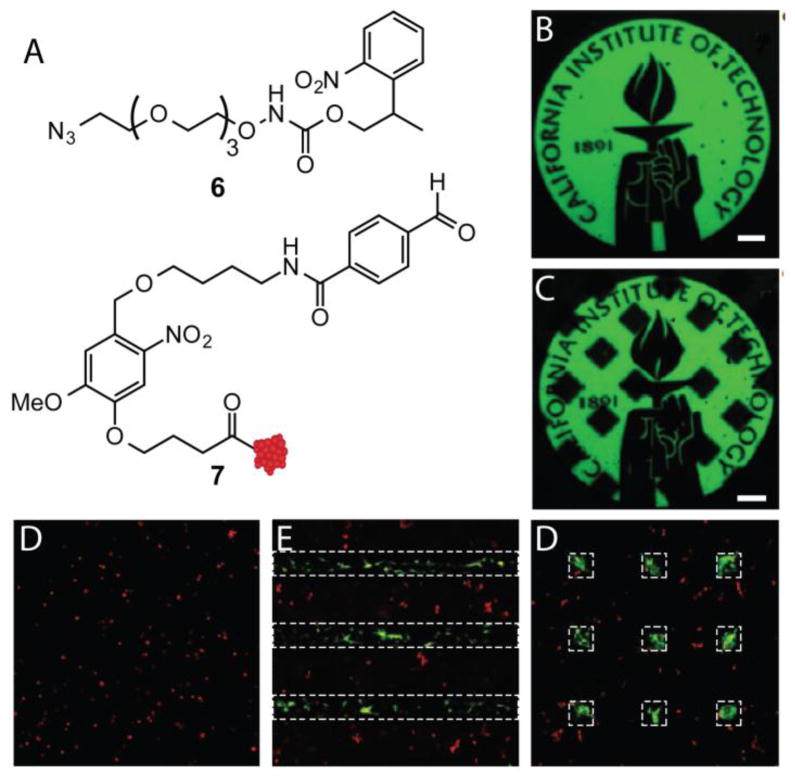
Figure 9.
SPAAC hydrogels were generated to bear masked alkoxyamine 6. Upon photolysis of ONB, biomolecules can be appended through the gel by aldehyde 7 that has an ONB linker to cleave biomolecules after bioconjugation. Fluorescent BSA resembling structure 7 was patterned within the hydrogel using multiphoton photolithography (B). At a later time point, fluorescent BSA was released from the hydrogel in defined regions (C) (Scale bars represent 100 μm). hMSCs were seeded within SPAAC hydrogels and treated with CellTracker red and stained for OC (green), a marker of osteogenesis. At 1 d, VTN was patterned in lines (shown as white dotted lines) and after 4 d, hMSCs stained for OC in patterned regions (E). VTN was released in regions after 4 d and hMSC OC expression was lost in those regions at 10 d (D).

