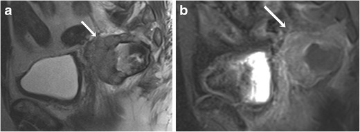
Fig. 8.

A 76-year-old male with anorectal melanoma. a Sagittal T2-weighted MR image shows a heterogeneous mass lesion arising from the anterior wall of the rectum (arrow). b The mass shows enhancement on the corresponding sagittal fat-suppressed T1-weighted MR image (arrow)
