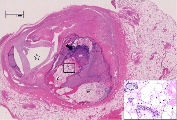
Fig. 1.

Coronary artery lesion in Kawasaki disease during follow-up. Extensive calcification (arrow) with ossification and bone marrow elements (insert, 400×) in the thrombosed and re-canalized left anterior descending artery from the explanted heart of a 29-year-old man who suffered from Kawasaki disease at age 3 years. The aneurysms remodelled, and the patient was discharged from follow-up at the age of 7 years when the coronary artery appeared normal by echocardiogram. The patient presented with progressive congestive symptoms at age 29 years and required cardiac transplantation. Characteristic ‘lotus root’ appearance of the artery results from thrombosis with recanalization (stars). Only one lumen remains patent (far left). Haematoxylin and eosin stain, 40×
