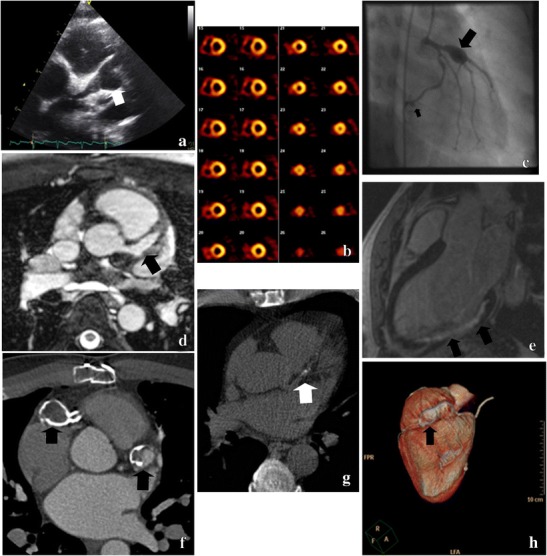
Fig. 2.

Imaging techniques for the follow-up of Kawasaki disease. a Echocardiogram of a giant aneurysm of the left main coronary artery (LMCA) and LAD. b Stress and rest SPECT Technetium-99M scan (myocardial perfusion scan) demonstrates ischemia of the inferior and septal wall. c Conventional CAG shows a giant aneurysm of the LAD and a smaller aneurysm of the right circumflex artery (RCX). d Cardiac MRI shows an aneurysm of the LAD. e Cardiac MRI indicates a myocardial infarction of the infero-posterior wall. f Multi-slice CT contrast-enhanced angiography with calcified aneurysms of the proximal LAD and right coronary artery (RCA). g CT calcium-score with calcifications of the proximal LAD. h 3D-CT angiography with a calcified aneurysm of the RCA
