Fig. 20.

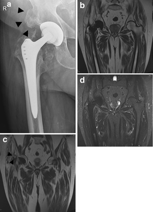
Although current state-of-the art MRI with Metal Artefact Reduction Sequences allows assessment of correct position of the hip prosthesis as well as periarticular abnormalities, mature heterotopic bone formation (arrowheads in A and C) is often more readily visible on plain radiographs than on MRI due to similar signal of mature bone marrow and fatty infiltration within the gluteus musculature at the site of the hip prothesis. a AP radiograph. Cementless total hip arthroplasty. Heterotopic bone formation (arrowheads), 7 years postoperatively. b T1-weighted, coronal image (WI) of the pelvis in the same patient. c T1-weighted, coronal image (WI) of the pelvis at a more anterior location barely showing heterotopic bone formation (arrowheads). d STIR, coronal image of the pelvis in the same patient
