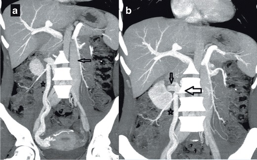
Fig. 5.

a Coronal MIP MDCT image showing left IVC (arrow) associated with dilated right gonadal vein (on the right) (asterisk) b Coronal MIP MDCT image showing RRV (small arrow) draining into the retroaortic left IVC (large arrow) in the paravertebral area, the dilated right gonadal vein is marked with an asterix
