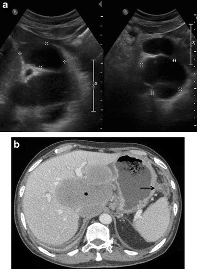
Fig. 1.

Incidentally detected PHL in a 76-year-old man with acute pancreatitis. a Gray-scale US images demonstrating a lobulated anechoic lesion (partly marked by calipers) in the gastrohepatic ligament abutting the liver. This was reported as suspicious for pancreatic pseudocysts in view of the clinical history. b Axial CECT image of the same patient showing a homogeneous, partially exophytic, hypoenhancing mass (asterisk) replacing the caudate lobe of the liver and involving the adjoining hepatic parenchyma. Biopsy from the liver lesion revealed findings consistent with diffuse large B-cell lymphoma. Sequel of acute pancreatitis can be seen as ill-defined soft tissue stranding along the greater curvature of the stomach (arrow)
