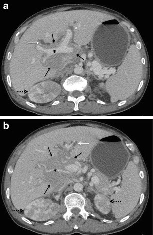
Fig. 15.

SHL in a 60-year-old man with jaundice. Axial CECT images demonstrating an ill-defined hypoenhancing periportal soft-tissue mass (solid black arrows in a and b) causing mild dilatation of the intrahepatic biliary radicals (white arrows). Portal vein and hepatic artery (asterisks in a and b, respectively) are seen coursing through the lesion without being attenuated or thrombosed. Bilateral kidneys are also involved (interrupted arrows)
