
Figure 1.
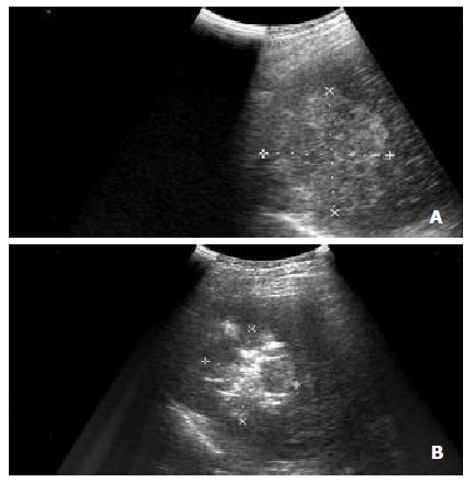
(A) Intercostal oblique sonogram shows a 5.7 cm diameter nodular HCC in segment 8. (B) The RF electrode has been placed in the lesion, and the electrode tip is recognizable as an echogenic area in the tumor. Sonogram obtained after several minutes of energy deposition shows a hyperechoic patch around the electrode tip.