
Figure 2.
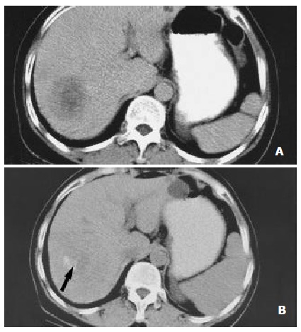
Nearly complete necrosis in a large HCC located in segment 7 and treated with RF therapy in a 57-year-old colorectal cancer liver metastases. (A) CT scan obtained prior to therapy demonstrates a large, 5.3 cm nodules. (B) CT scan obtained 6 mo after RF therapy shows nearly complete tumor necrosis. A small hypervascularized area of viable neoplastic tissue (arrow) remains at the posteromedial aspect of the tumor.