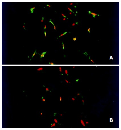
Figure 5.

Immunoflurescence detection of the expression of tyrosine phosphorylation in HSC by confocal laser microscopy (× 200). (A) HSC stimulated with 10 μg·L-1 PDGF-BB for 15 min showed a strong staining for phosphoyrosine containing protein; (B) HSCs stimulated with 10 μg·L-1 PDGF-BB for 15 min and then incubated with 10-7 mol/L genistein for 48 h showed significant decreased fluorescence intensity for phosphoyrosine containing protein.
