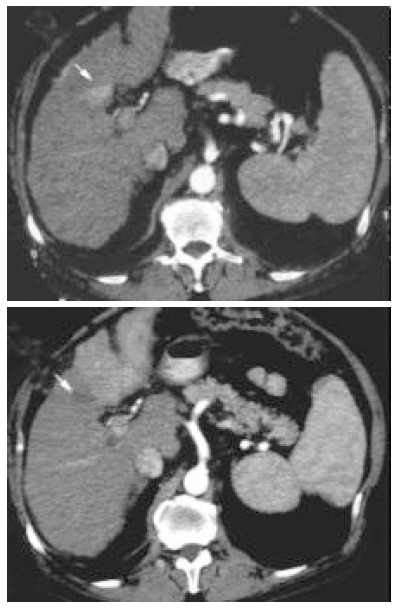
Figure 1.

Contrast-enhancement arterial phase computed tomography (CT) scans before treatment (upper image) and 30 d after treatment (lower image). Tumor shows local necrosis (arrow) and rapid progression of the tumor in the left lobewith pathologic enhancement at CT.
