
Figure 6.
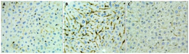
Immunohistochemical assay of ICAM-1 on liver tissue. Compared with sham control (A), the expression of ICAM-1 was highly up-regulated by 90 min of ischemia and 6 h of reperfusion (B), and the increase of ICAM-1 expression was dramatically inhibited by YS (20 mg/kg) (C). Original magnification: × 400.