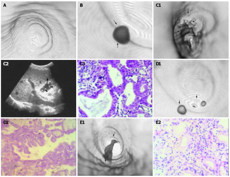
Figure 1.

CTVEGB detection of gallbladder polyps. A: Surface detail of gallbladder displayed by CTVEGB in a 47-year old normal man. B: Smallest single cholesterol polyp (arrow head) detected by CTVEGB in a 28-year-old man. C: Multiple gallbladder polyps in a 30- year-old man. (1) Multiple polyps of cauliflower appearance and small polyps (arrow head). (2) Color ultrasonography found multiple polyps of cauliflower appearance and small polyps (arrow head). (3) Multiple polyps were inflammatory polyps on pathology (HE × 20). D: Multiple cholesterol gallbladder polyps in a 30-year-old woman. Two cholesterol polyps (arrow head) were proved by pathology (HE × 20). E: Single inflammatory gallbladder polyps in a 29-year-old woman. An irregular inflamma-tory polyp was proved by pathology (HE × 20).
