
Figure 1.
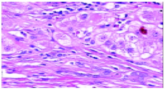
HLC. Several proliferating bile ductules are seen at the edge of a regenerating nodule. There is a dense, mostly lym-phocytic inflammatory infiltrate. Haematoxylin and eosin × 200.
Official websites use .gov
A
.gov website belongs to an official
government organization in the United States.
Secure .gov websites use HTTPS
A lock (
) or https:// means you've safely
connected to the .gov website. Share sensitive
information only on official, secure websites.
HLC. Several proliferating bile ductules are seen at the edge of a regenerating nodule. There is a dense, mostly lym-phocytic inflammatory infiltrate. Haematoxylin and eosin × 200.