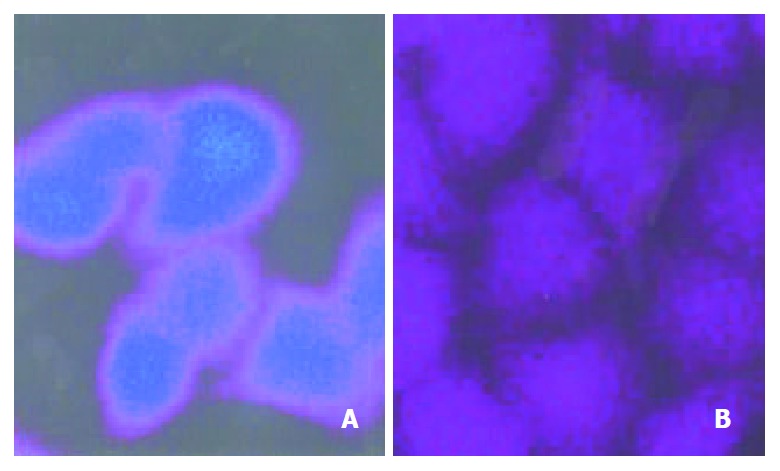
Figure 3.

FLU distribution in Bel7402 and Bel7402/5-FU cells. The cells were incubated with 100 μmol·L-1 FLU for 3 hr. Then the intracellular distribution of FLU was revealed by confocal la-ser scanning microscopy. (A) Bel7402 cells; (B) Bel7402/5-FU cells.
