
Figure 1.
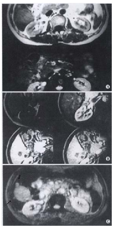
Primary hepatocellular carcinoma in posterior right lobe. The lesions appear hypointenity on T1WI and mildly hyperintensity on T2WI (A). On Gd-DTPA enhanced images, early enhancement can be seen in arterial phase and appear relatively hypointensity in portal phase.(B) On SPIO-enhanced image, the conspicuity is clearer than pre-contrasted and Gd-DTPA enhanced images. Two micro-lesions(arrow) are obviously showed (C).