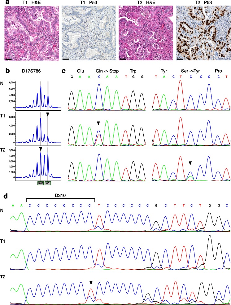
Fig. 2.
Routine genomic DNA and mitochondrial DNA results for patient 352, whom was diagnosed with synchronous tumors of the right (T1) and the left lung (T2). a Both tumors were diagnosed as adenocarcinomas with a bronchioloalveolar growth pattern; T1 shows absence of P53 staining, whereas T2 shows clear nuclear P53 staining. Scale bars represent 50 μm. b Routine genomic DNA analysis was performed on DNA isolated from normal (N) and both tumor tissues (T1 and T2). LOH analysis of marker D17S786 (TP53) showed loss of the large allele in T1 and loss of the small allele in T2, indicated by arrowheads. The horizontal axis indicates the size of the DNA fragments in basepair; the vertical axis indicates signal intensity. c Routine Sanger sequencing of TP53 showed a p.Gln52* mutation only in T1, and a p.Ser127Tyr mutation only in T2, both indicated by arrowheads. d Sanger sequencing of mitochondrial DNA marker D310 showed an 8-cytosine repeat in normal DNA, no aberrations in T1, and a 1-bp deletion in T2, as indicated by the arrowhead. The results of routine genomic DNA and mitochondrial DNA analysis both indicate that T1 and T2 represent two primary tumors. H&E hematoxylin and eosin stain

