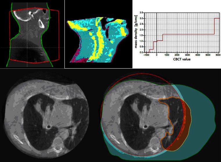
Fig. 1.
The RayStation treatment planning system (TPS) cone beam computed tomography (CBCT) CT number (CTN) adjustment method. Top row from left: Sagittal slice of a CBCT image of an H&N cancer patient viewed within the TPS; the CBCT after density assignment by the TPS (regions assigned as bone are shown as yellow, for example); CTN-density table generated for the CBCT image. Bottom row, left: Typical CBCT acquisition of a patient with a tumor in their right lung and (right) the same CBCT image but with the field of view (red contour), external (green contour), and left lung (orange contour) regions of interest displayed

