
Figure 2.
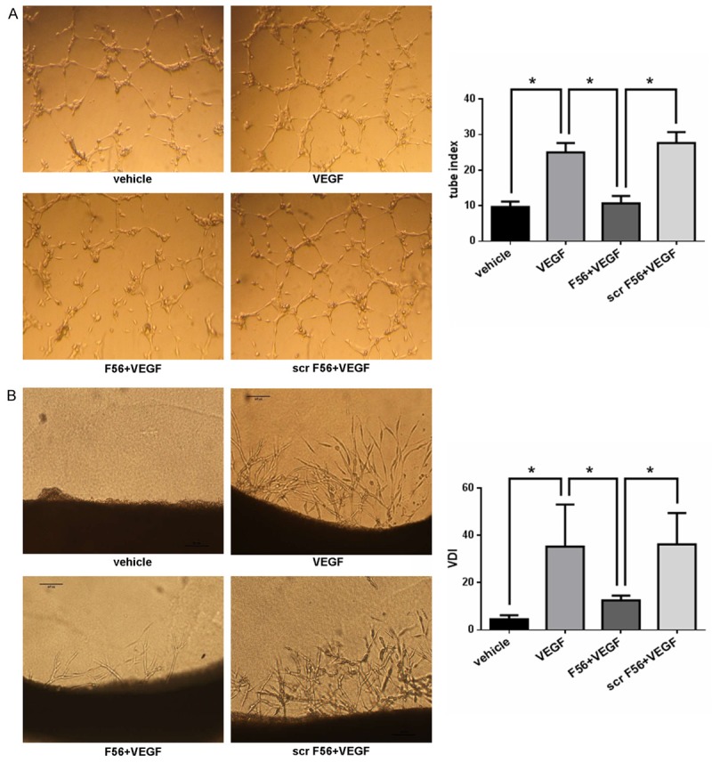
F56 inhibited VEGF induced HUVEC tube formation and endothelial cell sprouting in 3D rat aortic ring assay. A. HUVEC suspensions were pre-incubated with peptides for 30 min before exposure to VEGF (10 ng/ml). Each well with polymerized Matrigel was filled with 100 μl cell suspensions containing 20,000 cells. After 6 hr, the tubes were photographed and analysed. B. Rats were killed and the thoracic aortas were cut into 1 mm length segments and embedded in polymerized Matrigel with additional fresh liquid of 60 μl Matrigel. The plate was then incubated at 37°C for 3 hr to allow the Matrigel polymerized firmly, and then 0.5 ml serum free culture medium with agents as indicated was added, the media were changed every 2 days. After 6 days of incubation, microvessel-like structures sprouting from the aortic rings were photographed and analysed. Data are expressed as mean ± SEM. *P < 0.05.