
Figure 5.
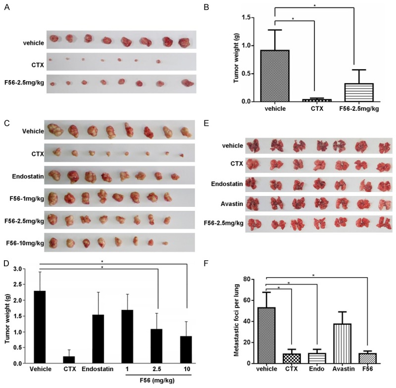
F56 inhibited tumor growth and metastasis in xenograft mice model. A. BALB/c nu/nu mice were implanted subcutaneously with 1 × 10⁷ HT-29 cells. After the tumor volume reached about 100 mm3, these mice were treated daily with vehicle or indicated drugs for consecutive 14 days. The tumor nodules were isolated one week after drug withdrawal. B. The weight of tumor nodules from A (n = 8). C. Tumor nodules formed by BGC-823 cells in the BALB/c nu/nu mice models were shown. D. The weight of tumor nodules from C (n = 8). E. C57 mice (n = 7) were injected with 4 × 10⁶ B16 melanoma cells via tail vein. Indicated drugs were delivered as described in the Methods. After continuous administration of drugs for 14 days, mice were killed, lungs were dissected and the number of metastatic foci per lung was counted carefully by naked eyes. F. The number of metastatic foci per lung from E was counted and analyzed. Data are expressed as mean ± SEM. *P < 0.05.