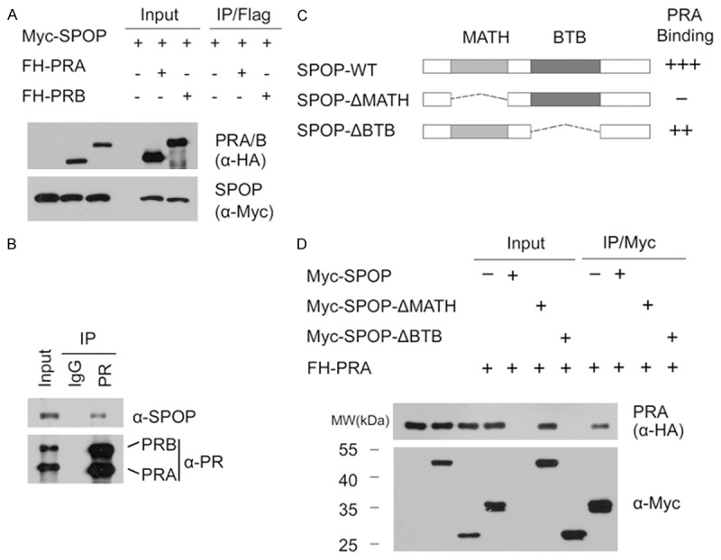
Figure 1.

SPOP interacts with PR in cells. A. 293T cells were co-transfected with Myc-SPOP and FH-PRA or PRB constructs. After 24 hr, cell lysates were prepared for co-IP with anti-Flag antibody and WB analyses; B. After treated with 20 µM MG132 for 4 hr, T47D cell lysates were prepared for co-IP with anti-PR antibody and WB analyses with indicated antibodies; C. Schematic representation of SPOP deletion mutants. Binding capacity of SPOP to PR is indicated with the symbol; D. 293T cells were co-transfected with FH-PRA and Myc-SPOP-WT or deletion mutants (ΔMATH, ΔBTB) constructs. After 24 hr, cell lysates were prepared for co-IP assay with anti-Myc antibody and WB analyses.
