. 2015 Oct 30;3(2):147–150. doi: 10.14791/btrt.2015.3.2.147

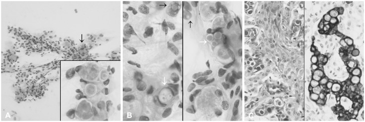
Fig. 2. Intraoperative crush and squash cytology of the resected mass. A: Cellular smear shows sheets of cohesive polygonal tumor cells forming occasional whorls (hematoxylin and eosin stain, ×200). Arrow and inset indicate eosinophilic globules (hematoxylin and eosin stain, ×1,000). B: Intracytoplasmic inclusions of variable size (white arrows) have a centrally located eosinophilic homogeneous core and a well-defined, thin, clear rim. Note frequent nuclear pseudoinclusions (black arrows, hematoxylin and eosin stain, ×1,000). C: Left: a resected mass is composed of large lobules of syncytially arranged ovoid shaped cells with periodic acid-Schiff-positive eosinophilic granular bodies in aggregated or singly scattered form. Note a periglobular thin halo-like rim (periodic acid-Schiff stain, ×400). Right: cells containing cytoplasmic secretory inclusions are occasionally positive for pancytokeratin (pancytokeratin immunostain, ×400).

Fig. 2