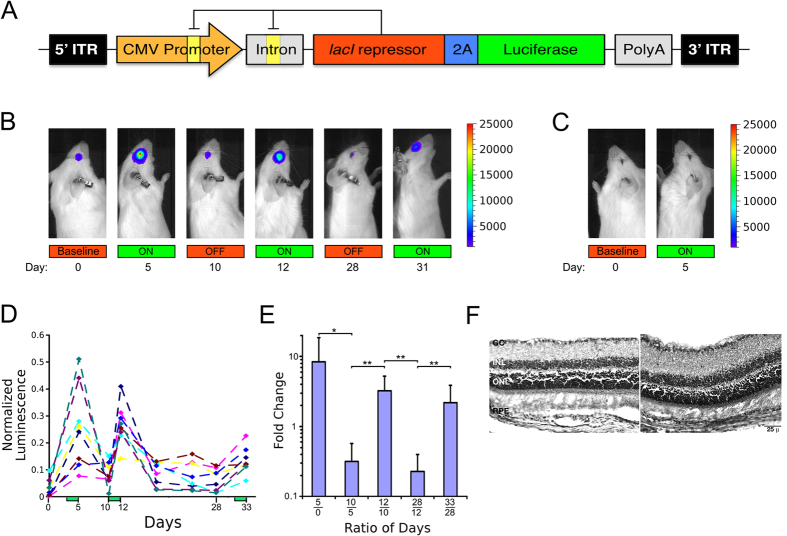
Figure 2. The autogenous regulatory system is functional in mouse retina in vivo.
(A) Map of the autogenously regulated expression system (ARES) within an AAV production vector (AAV8.ARES.Luciferase). A CMV promoter controls the expression of both the lacI repressor and Luciferase, linked via a 2A peptide cleavage sequence. Orange boxes indicate lac operator sites. Intronic, polyadenylation, and AAV ITR sequences are indicated. (B) Live imaging of luciferase activity over a 33-day period in a representative animal subretinally injected with AAV8.ARES.luciferase in the right eye. (C) Live imaging of luciferase activity in the left, un-injected eye of the same animal as in panel (A). (D) Normalized integrated luminescence of the injected (right) eye were calculated by dividing the observed luminescence by the sum of luminescent measurements made in both the on and off states for each animal. Induction of luciferase in AAV injected eye increases significantly after administration of IPTG (P < 0.01). Green bars represent days of IPTG gavage. (E) The fold change was determined by evaluating the normalized integrated luminescence on day n relative to day m, where n and m are labeled on the x-axis as  to illustrate dynamic regulation. A fold change >1 indicates induction of luciferase expression while fold change <1 indicates repression of luciferase expression. *p < 0.05, **p < 0.01, n = 8. (F) Histological sections of injected retinas from two representative animals stained with hematoxylin and eosin. (RPE, retinal pigmented epithelium, ONL, outer nuclear layer, INL, inner nuclear layer, GC, ganglion cell layer).

