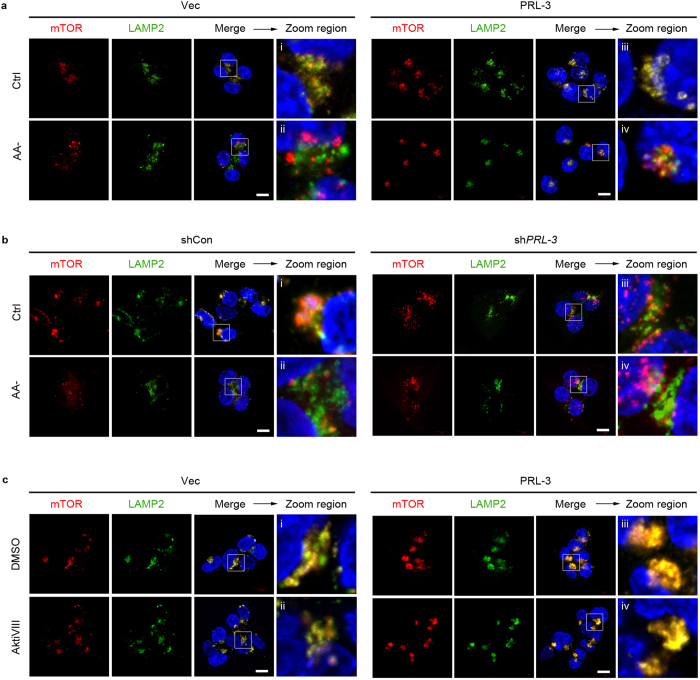
Figure 4. PRL-3 promotes the relocalization and accumulation of lysosomal mTOR in an Akt-independent manner.
(a) Immunofluorescence analysis of mTOR and LAMP2 in HCT116 cells overexpressing EGFP vector only (Vec) or EGFP-tagged wild-type PRL-3 (PRL-3) cultured in full media (Ctrl) or amino-acid starvation media (AA-). Antibodies against human LAMP2 and mTOR were used. Red, mTOR signal; green, LAMP2 signal; merge, merged mTOR, LAMP2, and DNA (DAPI) signals. Scale bar, 10 μm. A zoomed area within each merged panel enables better visualize mTOR/LAMP2 colocalization. (b) HCT116 cells stably expressing small hairpin RNA (shRNA) against PRL-3 (shPRL-3) or control shRNA (shCon) were cultured and analysed as in (a). Scale bar, 10 μm. (c) HCT116 Vec or PRL-3 cells were treated with DMSO or Akt Inhibitor VIII (AktiVIII) for 30 min and analyzed by dual immunfluoresence using antibodies against mTOR and LAMP2. Red, mTOR signal; green, LAMP2 Scale bar, 10 μm.

