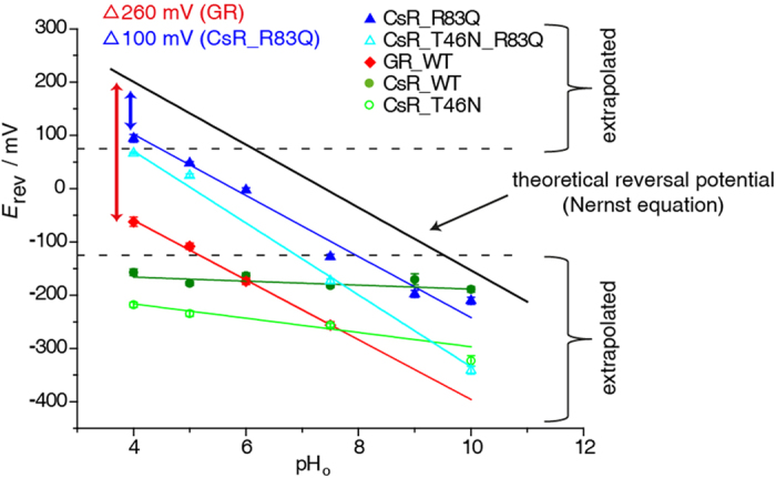
Figure 7. Reversal voltages of Δp- and ΔΨ-type light-driven proton pumps.

Reversal voltage (Erev) of Gloeobacter rhodopsin (GR), CsR-WT, CsR-T46N, CsR-R83Q, and CsR-R83Q-T46N are plotted against the extracellular pHo. The black line represents the theoretical expected Erev of free proton flow calculated by the Nernst equation based on an intracellular pHi of 7.4. Reversal potentials below this line indicate proton pump activity. Values from −125 mV up to +75 mV were determined directly. Other values are determined by extrapolation. CsR-WT (550 ± 25 nm, pH 10 [n = 8], pH 9 [n = 3], pH 7.5 [n = 26], pH 6 [n = 5], pH 5 [n = 8], pH 4 [n = 6]) exhibited high resistance against low pHo whereas GR (400–600 nm, pH 7.5 [n = 23], pH 6 [n = 6], pH 5 [n = 9], pH 4 [n = 10]) was shifted −260 mV in parallel to the calculated potentials. Mutation of CsR-R83 (550 ± 25 nm, pH 10 [n = 7], pH 9 [n = 4], pH 7.5 [n = 11], pH 6 [n = 4], pH 5 [n = 6], pH 4 [n = 4]) into Glu converts Coccomyxa rhodopsin into a Gloeobacter-like proton pump in relation to the Erev. CsR-T46N (550 ± 25 nm, pH 10 [n = 4], pH 7.5 [n = 8], pH 5 [n = 4], pH 4 [n = 4]). CsR-T46N-R83Q (550 ± 25 nm, pH 10 [n = 4], pH 7.5 [n = 5], pH 5 [n = 5], pH 4 [n = 2]). Data were measured in oocytes and represent the mean ± SE.
