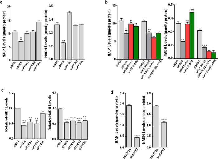
Figure 5. Blockade of proline biosynthesis downregulated NAD+ and NADH levels.
(a, b) PC9 lung cancer cells were transfected with siP5CS, individual siPYCR or all three siPYCRs simultaneously, and cultured under the indicated conditions for 4 ds. The levels of NAD+ and NADH were measured. (c, d) P493 lymphoma MYC tet-off cells were cultured under indicated conditions or transfected with siP5CS or individual siPYCR under MYC-On condition for 4 ds. NAD+ and NADH were measured. Data shown (mean ± S.E.M., n = 3) represent one of three independent experiments. P values were obtained by two-tailed Student’s t-test. *P < 0.05, **P < 0.01, ***P < 0.001 compared with the siNEG control or MYC-On group. #P < 0.05, ###P < 0.001 compared with the same knockdown (siP5CS) group in 5b.

