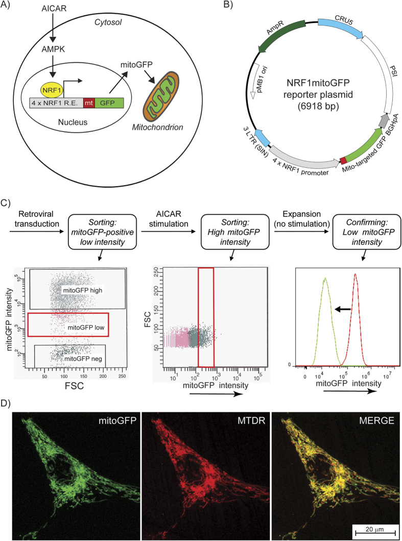
Figure 1. The live-cell mitochondrial reporter model.
(A) Overview of the live-cell mitochondrial reporter model. A reporter construct (NRF1mitoGFP) with mitoGFP under the control of a promoter with NRF-1 responsive elements (R.E.) was inserted in HeLa cells. AICAR was used to trigger NRF-1 reporter activity, via AMPK. The cellular expression of mitoGFP (intensity) reflected transcriptional responses of mitochondrial biogenesis, whereas the mitochondrial localisation of the mitoGFP protein enabled quantitative image analysis of mitochondrial morphology and biomass. (B) NRF1mitoGFP construct map. (C) Establishment of reporter cells. Flow cytometry was used to sort responding cells based on mitoGFP expression, depending on the presence of AICAR. The figure shows the stepwise strategy to isolate cells with inducible and reversible mitoGFP expression after treatment with AICAR (0.5 mM). The red gates in the two dot plots show the sorted populations. The histogram shows how the mitoGFP intensity shifted from high (red curve) to low (green curve) when AICAR was removed from the reporter cells. (D) Colocalisation of mitoGFP and MitoTracker Deep Red (MTDR) in HeLaNRF1/c4 cells after 6 days treatment with AICAR (0.5 mM). The images show contrast corrected maximum intensity composites (MICs) of a representative cell (imaged on a Zeiss LSM 510 META confocal microscope).

