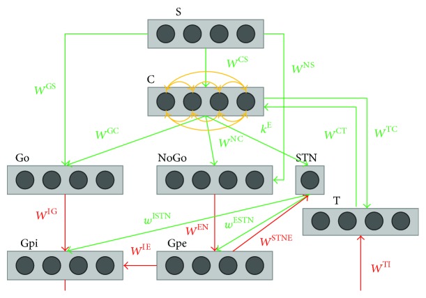
Figure 1.
Graphical representation of the overall basal ganglia model. Rectangles represent different structures, circles neurons, arrows projections: green excitatory, red inhibitory, and orange lateral inhibition. This figure depicts the principal areas taken into consideration in the current model: the sensory representation of the stimulus in the cortex (S), the motor representation in the cortex (C), the thalamus (T), the striatum, functionally divided according to dopamine receptor expression (Go and NoGo), the subthalamic nucleus (STN), the globus pallidus pars externa (Gpe), and the globus pallidus pars interna (Gpi). The synapses where learning takes place are those from the cortex to the Go (W GC) and the NoGo (W NC) part of the striatum and those from the stimulus representation S to the Go (W GS) and the NoGo (W NS) part of the striatum.

