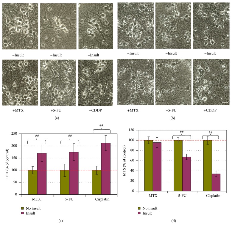
Figure 1.
Neurotoxicity of MTX, 5-FU, or CDDP in an AO-dependent manner. (a, b) Phase contrast photographs for neurons in 25% AO and 12.5% AO, respectively (bar = 25 μm). (c) Cell membrane leakage was determined via the LDH analysis at the end of experiment for neurons in 25% AO (n = 5). (d) Cell viability was determined with MTS analysis for neurons in 12.5% AO (n = 5). The bar in green and bars in other colors indicate the cultures without an insult and with anticancer drug insults, respectively. LDH and MTS data are expressed as % of noninsulted control. Figure symbol is ##, P < 0.01, compared with noninsult control group by Student's t-test.

