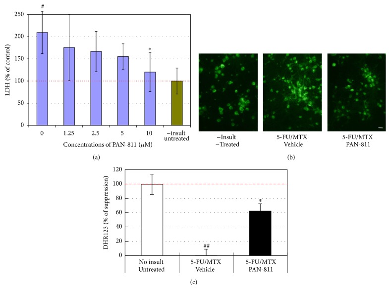
Figure 3.
Suppression of 5-FU/MTX-induced increases in LDH and DHR123 readings by PAN-811. (a) LDH release analysis for neurons that were cultured in 17.5% AOs-containing medium and insulted with both 100 μM MTX and 25 μM 5-FU in the absence or presence of PAN-811·Cl·H2O at different concentrations for 1 day (blue bars n = 6); Green bar represents noninsult/untreated control. (b) Fluorescent microscope for neurons in 17.5% AOs-containing medium insulted with both 100 μM MTX and 25 μM 5-FU in the absence or presence of 10 μM PAN-811·Cl·H2O for 1 day and incubated with DHR123 for 30 min (bar = 50 μm). (c) Quantification of (b) at excitation at 485 nm and emission at 520 nm (n = 4). Data are expressed as % suppression = [(Insulted&Untreated − Insulted&Treated)/(Insulted&Untreated − NonInsulted&Untreated) ∗ 100%]. Figure symbols are ∗, P < 0.05, compared with insult group alone (without PAN-811 treatment) by Student's t-test (one tail) and one-factor ANOVA followed by Tukey HSD test; #, P < 0.05; ##, P < 0.01, compared with noninsult/untreated control group by one-factor ANOVA followed with Tukey HSD test.

