Figure 7.
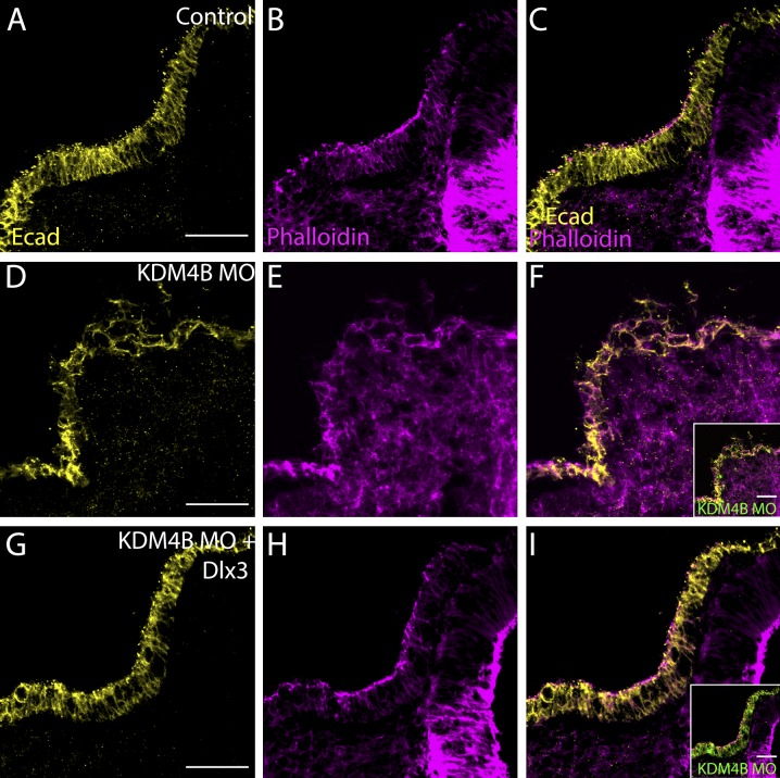
Coelectroporation of KDM4B-MO with pCIG-Dlx3 was sufficient to rescue otic invagination. Using immunohistochemistry, the control side (A–C) shows normal invagination as visualized by E-cad and phalloidin staining, but disrupted invagination on the KDM4B-MO–treated side (D–F). Coexpression of Dlx3 (G–I) with KDM4B-MO was sufficient to rescue otic invagination, leading to a proper arrangement of actin filaments and correct localization of E-cad. Bars, 50 µm.

