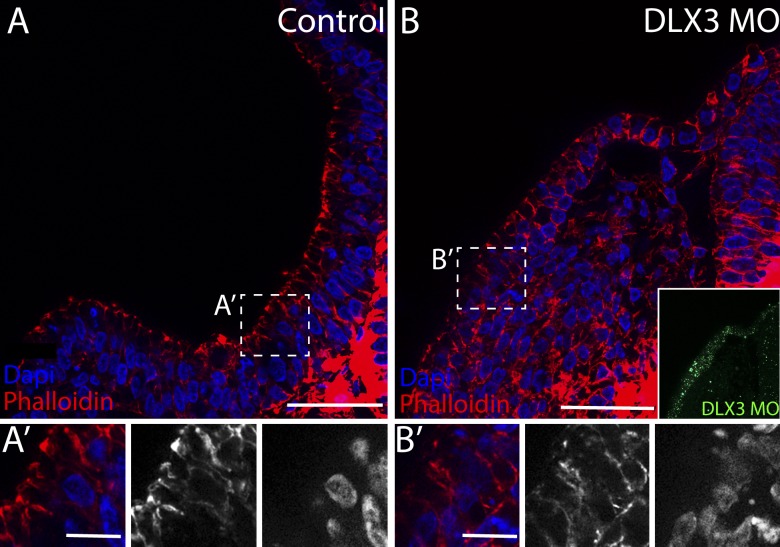
Figure 8.
Knockdown of Dlx3 results in failure of otic invagination. The control side (A) shows normal invagination as seen by apical accumulation of actin filaments viewed by phalloidin staining. The electroporation of DLX3-MO compromise otic invagination (B), indicated by irregular rearrangement of actin filaments. (A’ and B’) Inset views indicate the lack of apical accumulation of actin filaments on the injected side. Bars: (A and B) 60 µm; (A’ and B’) 20 µm.

