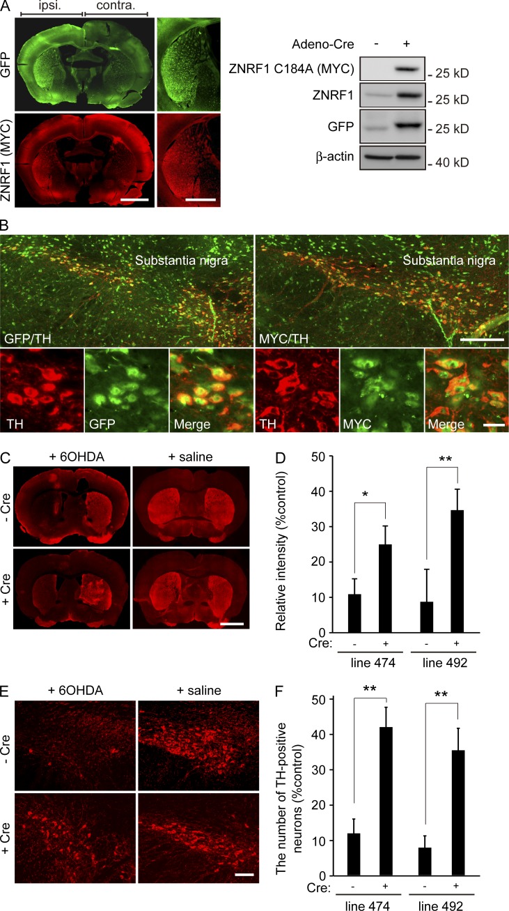
Figure 8.
Inhibition of ZNRF1 ubiquitin ligase activity preserves dopaminergic neurons in an in vivo 6OHDA-lesioned model. (A and B) A unilateral intranigral injection of the Cre-expressing adenoviral vector was performed to induce ZNRF1 C184A expression in the two independent Tg lines (lines 474 and 492) bearing ZNRF1 C184A–IRES–GFP cDNA, the expression of which is induced by the Cre-mediated excision of the loxP-flanked cassette. Transgene expression levels in the striatum (A, left) or SN (B) were confirmed by immunostaining using anti-GFP and anti-MYC antibodies. An immunoblot analysis against cell lysates from the ipsilateral (+) or contralateral (−) striatum 5 d after the adenovirus injection was also performed (A, right). Bars: (A) 500 µm; (B, top) 250 µm; (B, bottom) 25 µm. (C–F) The neuroprotective effects of ZNRF1 C184A expression were assessed by TH immunoreactivity 7 d after the 6OHDA injection into the striatum (C and D) and SN (E and F). Representative photomicrographs for the immunostaining of TH on coronal sections are shown in C. Bar, 500 µm. Relative immunofluorescent intensities in the ipsilateral to contralateral striatum for each condition are shown in D. Representative photomicrographs of the immunostaining of TH in the SN are shown in E. Bar, 250 µm. Ratios of the number of TH-positive neurons in the ipsilateral to contralateral SN for each condition are shown in F. Data are presented as the mean ± SEM. n = 3. Significant differences from the no infection condition (*, P < 0.05; **, P < 0.01) were determined by the two-tailed Student’s t test.

