Figure 1.
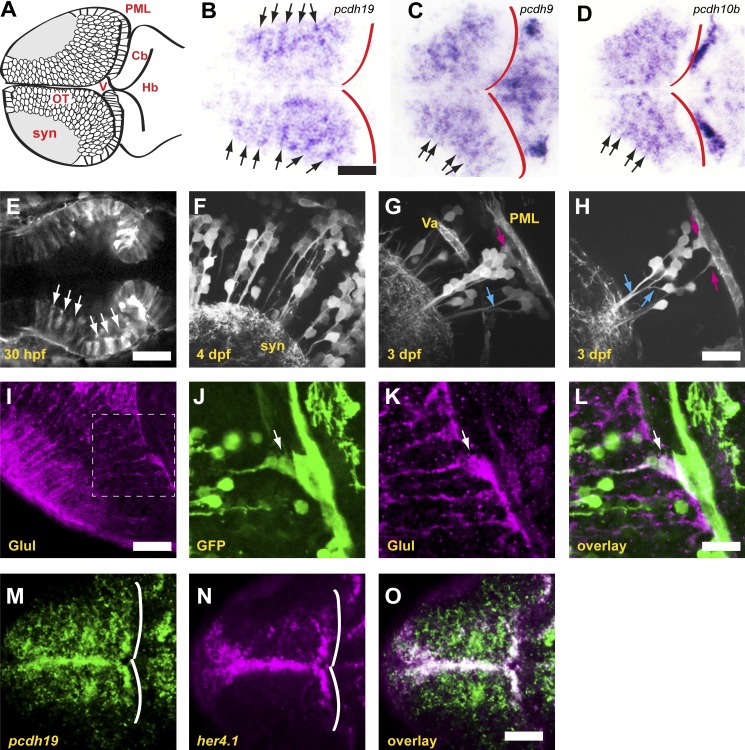
δ-Protocadherins define neuronal columns in the zebrafish optic tectum. (A) Schematic of the optic tectum of a larval zebrafish. Neurons are organized around a synaptic neuropil that includes both the axons and dendrites of tectal neurons, as well as the axonal arbors of retinal ganglion cells. Cb, cerebellum; Hb, hindbrain; OT, optic tectum; PML, peripheral midbrain layer; syn, synaptic neuropil; V, ventricle. (B) Horizontal section through the optic tectum of a 4-dpf larva labeled with a riboprobe against pcdh19. Labeling for pcdh19 is concentrated into radial stripes (arrows). The posterior boundary of the tectum is shown by red outlines. Bar, 50 µm in B–D. (C and D) Horizontal sections of optic tecta labeled with riboprobes against pcdh9 (C) and pcdh10b (D) showing radial stripes comparable to those observed for pcdh19. (E) Single optical section from a two-photon image stack of a 30-hpf BAC transgenic embryo expressing the F-actin marker, Lifeact-GFP, under the control of pcdh19 regulatory elements. The midbrain neuroepithelium expresses Lifeact-GFP in distinct stripes (arrows). Bar, 50 µm. (F) Maximum-intensity projection of five optical sections (1-µm spacing) of a 4-dpf BAC transgenic larva. Lifeact-GFP is expressed in discrete columns of neurons, as was observed by in situ hybridization for endogenous pcdh19. syn, synaptic neuropil. (G) Maximum-intensity projection of five optical sections (1-µm spacing) of a 3-dpf BAC transgenic larva. Labeled neurons are clustered around a central radial glia (magenta arrow), and most of the primary neurites fasciculate as they project to the synaptic neuropil (blue arrow). Va, vasculature; PML, posterior midbrain layer. (H) Single optical section through a column in a 3-dpf larva. Columns of pcdh19+ neurons are typically associated with one or more pcdh19+ radial glia (magenta arrows). Fasciculation of neuronal processes is highlighted by blue arrows. Neurons arborize in the area immediately adjacent to the column. Bar, 20 µm in F–H. (I–L) Horizontal sections through the optic tectum of a 4-dpf BAC transgenic larva, labeled with antibodies against glutamine synthase (Glul) to reveal radial glia (I and K) or GFP (J). The boxed area in I is highlighted in J–L. (L) Glutamine synthase staining reveals that a subset of radial glia are also pcdh19+ (arrow). Bars: (I) 30 µm; (J–L) 15 µm. (M–O) Single optical section through the dorsal optic tectum of a 3-dpf larva labeled by double FISH with riboprobes against pcdh19 (M) or the neural progenitor marker her4.1 (N). White lines show the posterior boundary of the tectum. An overlay of the signals reveals colocalization along the medial and posterior margins of the tectum (O). Bar, 50 µm.

