Figure 4.
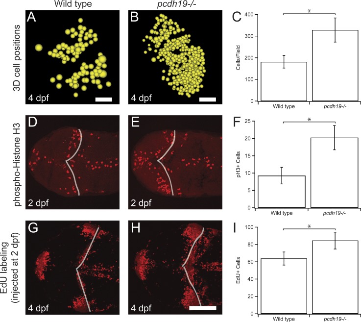
Increased proliferation in the optic tectum of pcdh19−/− mutants. (A–C) Two-photon image stacks were collected in WT (A) or pcdh19−/− (B) larvae. The 3D positions of transgenically labeled cell bodies were modeled as spheres. (C) The number of labeled cells was counted, confirming the visual impression of increased numbers of labeled cells in the pcdh19−/− mutants (WT, 181 ± 29, n = 6; pcdh19−/−, 328 ± 56, n = 5; *, P = 0.036, Student’s t test). Bars: (A) 20 µm; (B) 25 µm. (D–F) To determine whether rates of cell proliferation in the tectum are changed in the mutants, we performed pH3 immunocytochemistry at 2 dpf in both WT (D) and pcdh19−/− (E) larvae. (F) The number of pH3-labeled cells in the tectum was counted, revealing a significant increase in proliferation in the pcdh19−/− mutants (WT, 9.3 ± 2.4, n = 3; pcdh19−/−, 20.3 ± 3.5, n = 4; *, P = 0.04, Student’s t test). (G–I) To confirm our observations with pH3 labeling, we labeled WT (G) or pcdh19−/− (H) larvae with EdU at 2 dpf and fixed the larvae at 4 dpf. (I) The number of EdU-labeled cells in the tectum was counted, confirming a significant increase in proliferation in the pcdh19−/− mutants (WT, 63.8 ± 7.6, n = 4; pcdh19−/−, 84.5 ± 9.8, n = 4; P = 0.032, Student’s t test). Bar, 50 µm.

