Fig. 3.

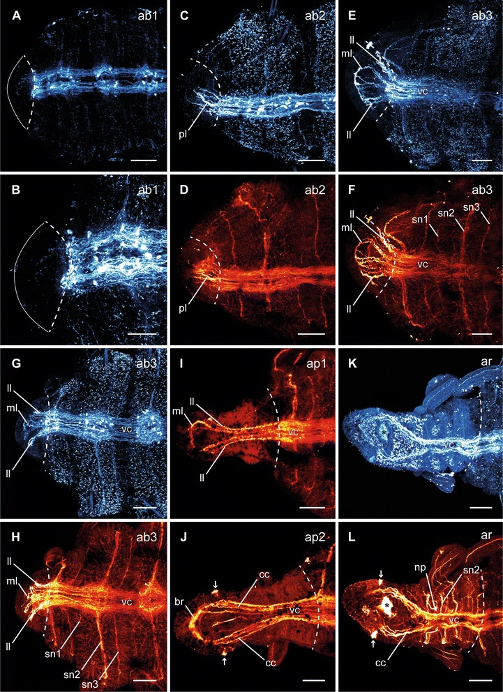
Different stages of anterior neuronal regeneration in Timarete cf. punctata. Anti-serotonin (cyan; A-C, E, G, K) and anti-acteylated α-tubulin (red; D, F, H-J, L) staining, confocal maximum projections. Anterior is left, all views are ventral showing the anterior end. The dotted white line indicates the side of dissection. Regeneration process is staged according to Fig. 2, time after decapitation of correspondent specimen is given in brackets. a, b Early blastema stage (ab1, 48 h). The nervous system has not been started to infiltrate the blastema (anterior margin indicated by white line). c, d Middle blastema stage (ab2, 68 h). Neurite bundles infiltrated the blastema, thereby forming a plexus (pl). e, f Late blastema stage (ab3, 92 h). The neurite bundles condensed into a three-loop-structure, composed of two lateral (ll) and one median loop (ml). The median loop (ml) is connecting the inner neurites of the ventral nerve cord (vc), whereas the lateral loops (ll) are solely connected to the outer neurites of their side. g, h Late blastema stage (ab3, 96 h). In this specimen the nerve loops (ll, ml) are more defined, but also bent more dorsal. i Early blastema patterning stage (ap1, 96 h). According to the elongation of the blastema, the nervous system was stretched anterior. Simultaneously, the transition of the nerve loops to the roots of the later circumesophageal connectives occured. While the median loop (ml) simply elongated, the lateral loops (ll) converged to one structure. j Late blastema patterning stage (ap2, 7 dad). The transition of the nerve loops to the circumesophageal connectives (cc) had finished. At the anterior end, the brain (br) had redeveloped. Also the nuchal organs (arrows) were visible, now. k, l Re-segmentation stage (ar, 11dad). Redevelopment of segmental nerves (sn2) according to reestablished segments was visible. Scale bars = 50 μm, except of A = 25 μm
